ARDMS SPI - ARDMS Sonography Principles and Instrumentation Certification Examination Exam
Question #1 (Topic: demo questions)
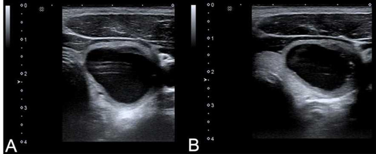
Which adjustment will reduce the artifact in the cystic lesion in image A resulting in image B?
Correct Answer: C
Explanation:
In image A, the borders of the cystic lesion might appear less defined due to a lack of edge enhancement. By turning on edge enhancement, the ultrasound system processes the image to accentuate the boundaries, leading to a clearer and more distinct outline of the cystic lesion as seen in image B. This adjustment reduces the artifact within the cystic lesion by emphasizing the differences in the adjacent tissue interfaces, thus improving the overall image quality. Reference: American Registry for Diagnostic Medical Sonography (ARDMS) Sonography Principles and Instrumentation guidelines on image optimization techniques.
Question #2 (Topic: demo questions)
Which target group is used to evaluate transverse distance measurement accuracy in this tissue- mimicking phantom image?
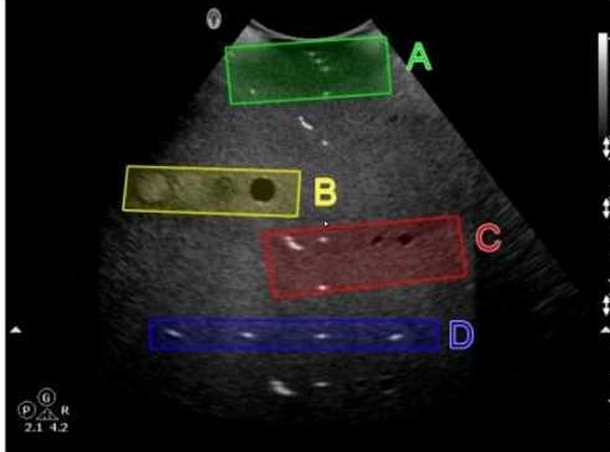
Correct Answer: D
Explanation:
In the tissue-mimicking phantom image, Option D (blue box) is used to evaluate transverse distance measurement accuracy. Phantoms are used to simulate human tissue and provide a standardized way to test the accuracy and precision of ultrasound machines. Transverse distance measurement accuracy is assessed by measuring known distances between targets in the phantom. The blue box (Option D) typically contains targets positioned to specifically test the accuracy of transverse measurements, ensuring that the ultrasound system provides reliable and precise distance readings. Reference ARDMS Sonography Principles and Instrumentation (SPI) Exam Study Guide "Quality Assurance for Ultrasound Imaging Systems" by AAPM (American Association of Physicists in Medicine)
Question #3 (Topic: demo questions)
Which artifact may be caused by incorrect color Dopplergain setting?
Correct Answer: A
Explanation:
Incorrect color Doppler gain settings can cause the artifact known as bleed or blossoming. When the color Doppler gain is set too high, it can cause the color signal to "bleed" outside the actual boundaries of the blood vessel, leading to an overestimation of the area of flow. This artifact makes it appear as though the blood flow extends beyond the true vessel walls, which can obscure the accurate interpretation of the Doppler image. Reference ARDMS Sonography Principles and Instrumentation (SPI) Exam Study Guide "Diagnostic Ultrasound: Principles and Instruments" by Frederick W. Kremkau
Question #4 (Topic: demo questions)
Which statement describes the purpose of using a spectral Doppler wall filter?
Correct Answer: D
Explanation: